Fish anatomy
From Wikipedia, the free encyclopedia

Contents
|
Fins

(1) - operculum (gill cover), (2) - lateral line, (3) - dorsal fin, (4) - fat fin, -- (5) - caudal peduncle, (6) - caudal fin, (7) - anal fin, (8) - photophores, -- (9) - pelvic fins (paired), (10) - pectoral fins (paired)
Spines and rays
In bony fish, most fins may have spines or rays. A fin can contain only spiny rays, only soft rays, or a combination of both. If both are present, the spiny rays are always anterior. Spines are generally stiff and sharp. Rays are generally soft, flexible, segmented, and may be branched. This segmentation of rays is the main difference that separates them from spines; spines may be flexible in certain species, but they will never be segmented.Spines have a variety of uses. In catfish, they are used as a form of defense; many catfish have the ability to lock their spines outwards. Triggerfish also use spines to lock themselves in crevices to prevent them being pulled out.
Types of fin
- Dorsal fins are located on the back. A fish can have up to three of them. The dorsal fins serve to protect the fish against rolling, and assists in sudden turns and stops.
- In anglerfish, the anterior of the dorsal fin is modified into an illicium and esca, a biological equivalent to a fishing pole and a lure.
- The bones that support the dorsal fin are called Pterygiophore. There are two to three of them: "proximal", "middle", and "distal". In spinous fins the distal is often fused to the middle, or not present at all.
- The caudal fin is the tail fin, located at the end of the caudal peduncle and is used for propulsion.
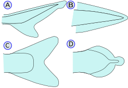
types of caudal fin :
(A) - Heterocercal, (B) - Protocercal,
(C) - Homocercal, (D) - Diphycercal- The tail can be heterocercal, which means that the vertebrae extend into a larger lobe of the tail or that the tail is asymmetrical
- Epicercal means that the upper lobe is longer (as in sharks)
- Hypocercal means that the lower lobe is longer (as in flying fish)
- Protocercal means that the caudal fin extends around the vertebral column, present in embryonic fish and hagfish. This is not to be confused with a caudal fin that has fused with the dorsal and anal fins to form a contiguous fin.
- Diphycercal refers to the special, three-lobed caudal fin of the coelacanth and lungfish where the vertebrae extend all the way to the end of the tail.
- Most fish have a homocercal tail, where the vertebrae do not extend into a lobe and the fin is more or less symmetrical. This can be expressed in a variety of shapes.
- The tail fin may be rounded at the end.
- The tail fin may be truncated, or end in a more-or-less vertical edge (such as in salmon).
- The fin may be forked, or end in two prongs.
- The tail fin may be emarginate, or with a slight inward curve.
- The tail fin may be lunate, or shaped like a crescent moon.
- The tail can be heterocercal, which means that the vertebrae extend into a larger lobe of the tail or that the tail is asymmetrical
- The anal fin is located on the ventral surface behind the anus. This fin is used to stabilize the fish while swimming.
- The paired pectoral fins are located on each side, usually just behind the operculum, and are homologous to the forelimbs of tetrapods.
- A peculiar function of pectoral fins, highly developed in some fish, is the creation of the dynamic lifting force that assists some fish, such as sharks, in maintaining depth and also enables the "flight" for flying fish.

Bigeye tuna Thunnus obesus showing finlets and keels.
Drawing by Dr Tony Ayling - In many fish, the pectoral fins aid in walking, especially in the lobe-like fins of some anglerfish and in the mudskipper.
- Certain rays of the pectoral fins may be adapted into finger-like projections, such as in sea robins and flying gurnards.
- The "horns" of manta rays and their relatives are called cephalic fins; this is actually a modification of the anterior portion of the pectoral fin.
- A peculiar function of pectoral fins, highly developed in some fish, is the creation of the dynamic lifting force that assists some fish, such as sharks, in maintaining depth and also enables the "flight" for flying fish.
- The paired pelvic or ventral fins are located ventrally below the pectoral fins. They are homologous to the hindlimbs of tetrapods. The pelvic fin assists the fish in going up or down through the water, turning sharply, and stopping quickly.
- In gobies, the pelvic fins are often fused into a single sucker disk. This can be used to attach to objects.
- The adipose fin is a soft, fleshy fin found on the back behind the dorsal fin and just forward of the caudal fin. It is absent in many fish families, but is found in Salmonidae, characins and catfishes.
- Some types of fast-swimming fish have a horizontal caudal keel just forward of the tail fin. This is a lateral ridge on the caudal peduncle, usually composed of scutes (see below), that provides stability and support to the caudal fin. There may be a single paired keel, one on each side, or two pairs above and below.
- Finlets are small fins, generally behind the dorsal and anal fins (in bichirs, there are only finlets on the dorsal surface and no dorsal fin). In some fish such as tuna or sauries, they are rayless, non-retractable, and found between the last dorsal and/or anal fin and the caudal fin.


Reproductive system
Internal fertilization
In many species of fish, fins have been modified to allow internal fertilization.A gonopodium is an anal fin that is modified into an intromittent organ in males of certain species of live-bearing fish in the families Anablepidae and Poeciliidae. It is movable and used to impregnate females during mating. The male's anal fin’s 3rd, 4th and 5th rays are formed into a tube like structure in which the sperm of the fish is ejected. In some species, the gonopodium may be as much as 50% of the total body length. Occasionally the fin is too long to be used, as in the "lyretail" breeds of Xiphophorus helleri. Hormone treated females may develop gonopodia. These are useless for breeding. One finds similar organs having the same characteristics in other types of fish, for example the andropodium in the Hemirhamphodon or in the Goodeidae.
When ready for mating, the gonopodium becomes “erect” and points forward, towards the female. The male shortly inserts the organ into the sex opening of the female, with hook-like adaptations that allow the fish to grip onto the female to insure impregnation. If a female remains stationary and her partner contacts her vent with his gonopodium, she is fertilized. The sperm is preserved in the female's oviduct. This allows females to, at any time, fertilize themselves without further assistance of males.
Male cartilaginous fish have claspers modified from pelvic fins. These are intromittent organs, used to channel semen into the female's cloaca during copulation.
Skin
See also: Scale (zoology)
The outer body of many fish is covered with scales. Some species are covered instead by scutes. Others have no outer covering on the skin; these are called naked fish. Most fish are covered in a protective layer of slime (mucus).There are four types of fish scales.
- Placoid scales, also called dermal denticles, are similar to teeth in that they are made of dentin covered by enamel. They are typical of sharks and rays.
- Ganoid scales are flat, basal-looking scales that cover a fish body with little overlapping. They are typical of gar and bichirs.
- Cycloid scales are small oval-shaped scales with growth rings. Bowfin and remora have cycloid scales.
- Ctenoid scales are similar to the cycloid scales, with growth rings. They are distinguished by spines that cover one edge. Halibut have this type of scale.
- an external shield-like bony plate, or
- a modified, thickened scale that often is keeled or spiny, or
- a projecting, modified (rough and strongly ridged) scale, usually associated with the lateral line, or on the caudal peduncle forming caudal keels, or along the ventral profile. Some fish, such as pineconefish, are completely or partially covered in scutes.
Vertebrae
The vertebrae of lobe-finned fishes consist of three discrete bony elements. The vertebral arch surrounds the spinal cord, and is of broadly similar form to that found in most other vertebrates. Just beneath the arch lies a small plate-like pleurocentrum, which protects the upper surface of the notochord, and below that, a larger arch-shaped intercentrum to protect the lower border. Both of these structures are embedded within a single cylindrical mass of cartilage. A similar arrangement was found in primitive tetrapods, but, in the evolutionary line that led to reptiles (and hence, also to mammals and birds), the intercentrum became partially or wholly replaced by an enlarged pleurocentrum, which in turn became the bony vertebral body.In most ray-finned fishes, including all teleosts, these two structures are fused with, and embedded within, a solid piece of bone superficially resembling the vertebral body of mammals. In living amphibians, there is simply a cylindrical piece of bone below the vertebral arch, with no trace of the separate elements present in the early tetrapods.
In cartilagenous fish, such as sharks, the vertebrae consist of two cartilagenous tubes. The upper tube is formed from the vertebral arches, but also includes additional cartilagenous structures filling in the gaps between the vertebrae, and so enclosing the spinal cord in an essentially continuous sheath. The lower tube surrounds the notochord, and has a complex structure, often including multiple layers of calcification.
Lampreys have vertebral arches, but nothing resembling the vertebral bodies found in all higher vertebrates. Even the arches are discontinuous, consisting of separate pieces of arch-shaped cartilage around the spinal cord in most parts of the body, changing to long strips of cartilage above and below in the tail region. Hagfishes lack a true vertebral column, and are therefore not properly considered vertebrates, but a few tiny neural arches are present in the tail
The jaw
See also: Wrasse#Anatomy


It is thought that the original selective advantage garnered by the jaw was not related to feeding, but to increased respiration efficiency. The jaws were used in the buccal pump (observable in modern fish and amphibians) that pumps water across the gills of fish or air into the lungs in the case of amphibians. Over evolutionary time the more familiar use of jaws (to humans), in feeding, was selected for and became a very important function in vertebrates.
Internal organs

- The gas bladder, or swim bladder, is an internal organ that contributes to the ability of a fish to control its buoyancy, and thus to stay at the current water depth, ascend, or descend without having to waste energy in swimming. It is often absent in fast swimming fishes such as the tuna and mackerel families.
- Certain groups of fish have modifications to allow them to hear, such as the Weberian apparatus of Ostariophysians.
- The gills, located under the operculum, are a respiratory organ for the extraction of oxygen from water and for the excretion of carbon dioxide. They are not usually visible, but can be seen in some species, such as the frilled shark.
- The labyrinth organ of Anabantoidei and Clariidae is used to allow the fish to extract oxygen from the air.
- Gill rakers are bony or cartilaginous, finger-like projections off the gill arch which function in filter-feeders in retaining food organisms.
- Electric fish are able to produce electric fields by modified muscles in their body.
- Many fish species are hermaphrodites. Synchronous hermaphrodites possess both ovaries and testes at the same time. Sequential hermaphrodites have both types of tissue in their gonads, with one type being predominant while the fish belongs to the corresponding gender.
- The blood circulation of fishes is called "single circuit circulatory system"
No comments:
Post a Comment
Hope you enjoyed your visit. Leave a comment and let us know. If you wish to ask a question please do so at the following link: http://tanks4thememories.blogspot.com/p/dear-tanks-advice-faq-column.html